| How to Learn in 24 Hours? |
|
| Need Help? |
M-F: 9am-5pm(PST):
Toll-Free: (877) RAPID-10
or 1-877-727-4310
24/7 Online Technical Support:
The Rapid Support Center
vip@rapidlearningcenter.com
Secure Online Order:

|
| Tell-A-Friend: |
Have friends taking science and math courses too? Tell them about our rapid learning system.
|
|
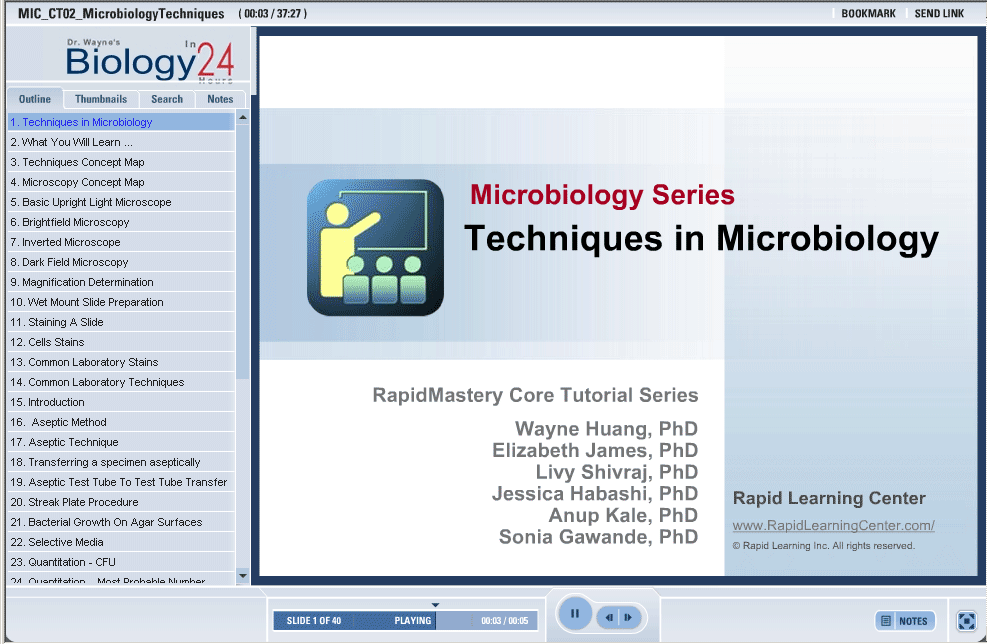
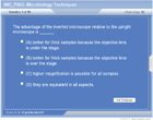
Techniques in Microbiology
| Topic Review on "Title": |
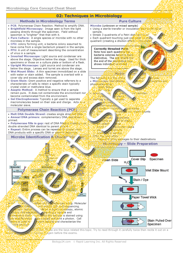
Microscopy:
- Electron Microscopy: uses electrons to create an image of the specimen.
- Brightfield Microscopy: specimen is visualized after the light has passed through it.
- Dark Field Microscopy: used when staining a sample is difficult or impossible.
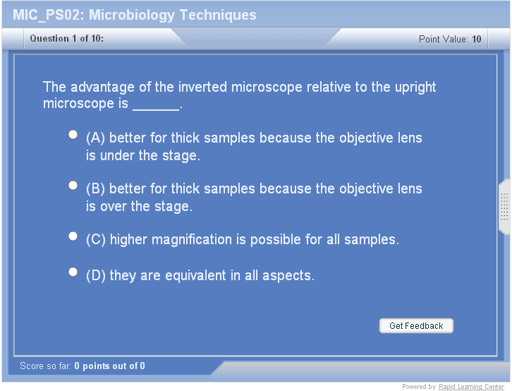
- Inverted Microscope: lenses are under the stage point upward used for thick samples.
- Upright Light Microscope: lenses are above the specimen point down.
Slide Preparation and Cell Stains:
- Wet Mount slide preparation: can be done with specific dyes or water.
- Common Laboratory Stains: Gram-Negative stain with safranin and appear red.
- Gram-Positive: crystal violet sometimes methylene blue.
Aseptic Techniques: Method to collect, grow and preserve a sample such that no other microbes are introduced or lost from the original culture.
Agar Plates:
- Streak Plate Procedure: plate is streaked in a zig zag patter to dilute a sample spread out sample for the purpose of isolating individual colonies.
- Selective Agar: growth medium with chemicals mixed in to select bacteria based on specific physiological characteristics e.g. antibiotic resistance.
- Quantification: Colony forming units (CFU) method to determine the number of bacteria in ample.
- Dilution Plating: successive dilution of a sample to quantify and isolate single colonies.
- Isolation of pure bacteria colonies: method to create a pure culture.
- Colony Characteristics: Shape, size, morphology and growth patterns.
- Phage Quantification: PFU, plaque forming units, clear areas of growth on a bacterial lawn plated on agar which indicate the presence and numbers of phage present.
DNA Methods
- PCR: Polymerase Chain Reaction, method used to amplify specific DNA sequences for ease of identification and sequencing.
- Electrophoresis: DNA sample place in a gel substrate between two electrodes. The charge and size of the DNA determines how fast the DNA migrates in the electrical field.
- DNA sequencing: Method to determine the order of which nucleotides are arranged on a strand of DNA.
|
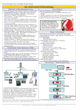
| Rapid Study Kit for "Title": |
| Flash Movie |
Flash Game |
Flash Card |
| Core Concept Tutorial |
Problem Solving Drill |
Review Cheat Sheet |
 |
 |
 |
|
| "Title" Tutorial Summary : |
This module introduces standard microbiology techniques utilized in laboratories. The techniques introduced are based on optical, molecular and biochemical principles. All of the methods involve use attributes of the microbe’s structural features.
The basic structural attributes of a microbe dictate the types of methods used to detect it and determine its identity. These methods can also be used to determine the physiology of the microbe, what its energy requirements are for growth and replication as well as any special attributes like antibiotic resistance it may have.
|
| Tutorial Features: |
Specific Tutorial Features:
- Microscopes, including light, electron and fluorescent are presented and their individual applications described.
- Step by step instruction on how to prepare slides for use with a light microscope is described.
- You will learn how to make pure bacterial and viral cultures.
- Molecular tools used in microbiology: PCR, electrophoresis and DNA sequencing.
- Systematic methods for quantifying bacteria and virus in a culture are taught.
- Aseptic methods to retain, isolate and grow pure cultures are introduced.
Series Features:
- Concept map showing inter-connections of concepts.
- Definition slides introduce terms as they are needed.
- Examples given throughout to illustrate how the concepts apply.
- A concise summary is given at the conclusion of the tutorial.
|
| "Title" Topic List: |
Microscopes:
Light,
Upright Microscopes
Brightfield Microscopy
Inverted Microscopy
Electron: Scanning Electron Microscopy
Fluorescent
Immune
Slide staining
Slide preparation
Cell Staining for Microbe Identification
Aseptic Techniques
Agar plate preparation and streaking for the purpose of individual colony isolation
Bacterial Growth on selective agar.
Quantification: Colony Forming Units (CFU)
Dilution Plating
Identification and characteristics of colonies.
PFU: plaque forming units for phage identification.
Bacteria Cultivation and Isolation
Selective Bacteria Growth Methods on Agar Surfaces
Quantitative Methods of Microbiology
Application and Analysis of PCR
|
See all 24 lessons in Anatomy and Physiology, including concept tutorials, problem drills and cheat sheets:
Teach Yourself Microbiology Visually in 24 Hours
|